|
This is a continuation of the article by Janina Radzikowska, Senior Metallographer, The Foundary Research Institute (Instytut Odlewnictwa) Kraków, Poland . It was originally published by Buehler in Tech-Notes, Volume 2, Issue 2. It is reproduced here with the kind permission of the Editor, Mr. Vander Voort who granted it while still with Buehler. |
 500x 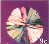 800x |
 500x 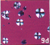 250x |
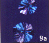
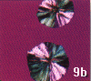
The polarizer and analyzer filters are placed in the crossed position (i.e., 90° to each other which produces the darkest matrix appearance), and a sensitive tint plate (lambda plate) may be inserted to further enhance coloration.The lambda plate may be adjustable on some microscopes which changes the color pattern, as illustrated in Figures 9a and b. Personally, I prefer to set the background color to magenta, as in Figure 9b. Working at a very high magnification, Figure 9c, provides more structural detail than at low magnification, Figure 9d, but requires high quality, stress- free objectives. Note the classic cross pattern seen at low magnification, Figure 9d. |
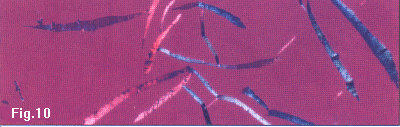
| Flake graphite can also be studied using polarized light, such as illustrated in Figure 10. The color varies with the crystallographic orientation of the graphite, and some internal details of the flake structure are better revealed in color. |
|
| Go to: | [FAQ] | [Home] | [Beginning of Article] |